International Mass Spectrometry Conference, Florence, Italy, August 28, 2018
Louisiana State University: Kermit K. Murray, Fabrizio Donnarumma, Kelin Wang, Carson W. Szot, Chao Dong,
Baylor University: Touradj Solouki, Michael E. Pettit
Matrix-assisted laser desorption/ionization (MALDI) mass spectrometry imaging (MSI) is a powerful method for determining the location of biomolecules in tissue; however, protein identification and quantification remains challenging. The goal of this project is to develop an imaging workflow that combines MALDI imaging with laser ablation microsampling for liquid chromatography tandem mass spectrometry.

Introduction
Matrix-assisted laser desorption/ionization (MALDI) mass spectrometry imaging (MSI) is a powerful method for determining the location of biomolecules in tissue; however, protein identification and quantification remains challenging. The goal of this project is to develop an imaging workflow that combines MALDI imaging with laser ablation microsampling for liquid chromatography tandem mass spectrometry.
Methods
In the combined workflow, MALDI imaging is used to identify regions of interest (ROI) from intact proteins. The ROI are sampled using infrared laser ablation and the captured material is analyzed by LC-MS/MS using data independent acquisition to identify and quantify the proteins. The data are cross-correlated to identify the localized proteins in the MALDI images.
Results
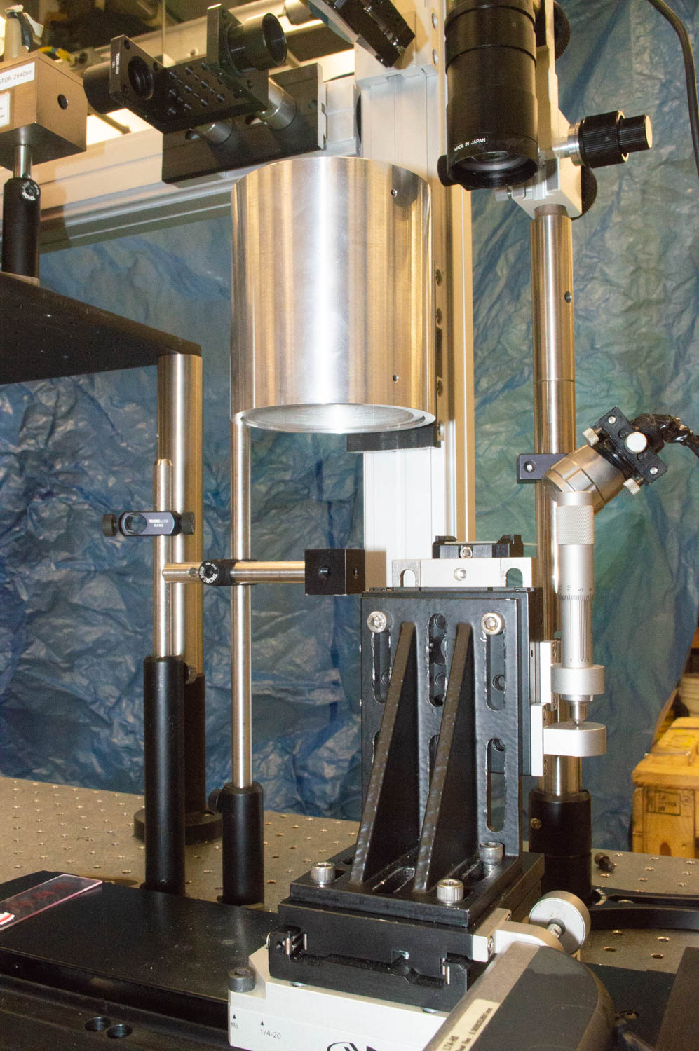
Development of the combined approach is aimed at creating an automated system for ablation and capture and using it in a coupled workflow of MALDI imaging and LC MS/MS analysis. The infrared laser ablation and capture system uses a mid-infrared optical parametric oscillator laser with a custom reflective objective that has a large working distance and good numerical aperture. We have developed custom positioning software that allows MALDI MSI heat maps to be overlaid on camera images to co-register ROI ablation with the IR laser. Tissue sections are mounted on conductive microscope slides and either consecutive sections or MALDI analyzed sections can be used. Laser ablated proteins are digested with magnetic capture beads and the peptides released for analysis with a Waters nanoAcquity UPLC system coupled to a Synapt G2-HDMS.
Conclusions
MALDI MSI coupled with region-specific laser ablation sampling for LC MS/MS is a fast and versatile approach for spatially resolved tissue proteomics. We have demonstrated that proteins can be identified from spatially localized regions and are developing new methods for correlating the intact proteins observed in MALDI with the proteins identified by tandem mass spectrometry.
Novel Aspect: Coupled MALDI imaging with high precision infrared laser ablation capture for LC MS/MS for protein identification and quantification.
International Mass Spectrometry Conference, Florence, Italy, August 28, 2018
Louisiana State University: Kermit K. Murray, Fabrizio Donnarumma, Kelin Wang, Carson W. Szot, Chao Dong,
Baylor University: Touradj Solouki, Michael E. Pettit
Introduction
Matrix-assisted laser desorption/ionization (MALDI) mass spectrometry imaging (MSI) is a powerful method for determining the location of biomolecules in tissue; however, protein identification and quantification remains challenging. The goal of this project is to develop an imaging workflow that combines MALDI imaging with laser ablation microsampling for liquid chromatography tandem mass spectrometry.
Methods
In the combined workflow, MALDI imaging is used to identify regions of interest (ROI) from intact proteins. The ROI are sampled using infrared laser ablation and the captured material is analyzed by LC-MS/MS using data independent acquisition to identify and quantify the proteins. The data are cross-correlated to identify the localized proteins in the MALDI images.
Results
Development of the combined approach is aimed at creating an automated system for ablation and capture and using it in a coupled workflow of MALDI imaging and LC MS/MS analysis. The infrared laser ablation and capture system uses a mid-infrared optical parametric oscillator laser with a custom reflective objective that has a large working distance and good numerical aperture. We have developed custom positioning software that allows MALDI MSI heat maps to be overlaid on camera images to co-register ROI ablation with the IR laser. Tissue sections are mounted on conductive microscope slides and either consecutive sections or MALDI analyzed sections can be used. Laser ablated proteins are digested with magnetic capture beads and the peptides released for analysis with a Waters nanoAcquity UPLC system coupled to a Synapt G2-HDMS.
Conclusions
MALDI MSI coupled with region-specific laser ablation sampling for LC MS/MS is a fast and versatile approach for spatially resolved tissue proteomics. We have demonstrated that proteins can be identified from spatially localized regions and are developing new methods for correlating the intact proteins observed in MALDI with the proteins identified by tandem mass spectrometry.
Novel Aspect: Coupled MALDI imaging with high precision infrared laser ablation capture for LC MS/MS for protein identification and quantification.