R.O. Lawal, F. Donnarumma, K.K. Murray, Deep-ultraviolet laser ablation electrospray ionization mass spectrometry, J. Mass Spectrom.54 (2019) 281–287. doi:10.1002/jms.4338.
Abstract

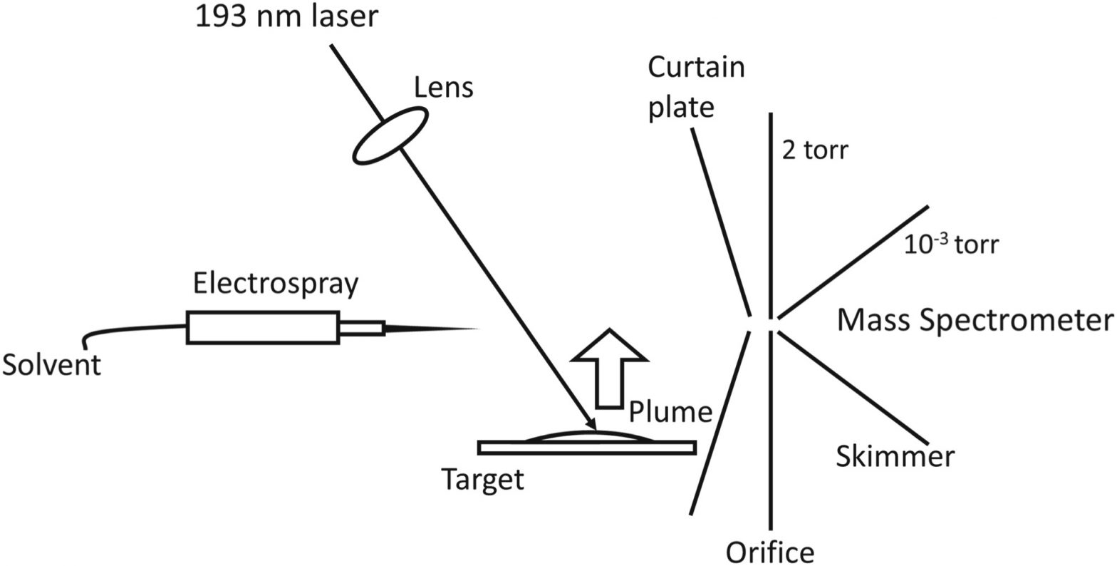
A 193‐nm wavelength deep ultraviolet laser was used for ambient laser ablation electrospray ionization mass spectrometry of biological samples. A pulsed ArF excimer laser was used to ablate solid samples, and the resulting plume of the desorbed material merged with charged electrospray droplets to form ions that were detected with a quadrupole time‐of‐flight mass spectrometer. Solutions containing peptide and protein standards up to 66‐kDa molecular weight were deposited on a metal target, dried, and analyzed. No fragmentation was observed from peptides and proteins as well as from the more easily fragmented vitamin B12 molecule. The mass spectra contained peaks from multiply charged ions that were identical to conventional electrospray. Deep UV laser ablation of tissue allowed detection of lipids from untreated tissue. The mechanism of ionization is postulated to involve absorption of laser energy by a fraction of the analyte molecules that act as a sacrificial matrix or by residual water in the sample.