B. Banstola, K.K. Murray, “A Nanoparticle Co-matrix for Multiple Charging in MALDI Imaging of Tissue,” Rapid Commun. Mass Spectrom. 16 (2019) 12. doi:10.1002/rcm.8424.
Abstract
Rationale: A two‐component matrix of 2‐nitrophloroglucinol (2‐NPG) and silica nanoparticles was used for matrix‐assisted laser desorption ionization (MALDI) mass spectrometry imaging of high‐charge‐state biomolecules in tissue. Potential advantages include increased effective mass range and efficiency of fragmentation.
Methods: A mixture of 2‐NPG matrix and silica nanoparticles was applied to cyrosectioned 10 μm thick mouse brain tissue. The mixture was pipetted onto the tissue for profiling and sprayed for tissue imaging. MALDI images were obtained under high vacuum in a commercial time‐of‐flight mass spectrometer.
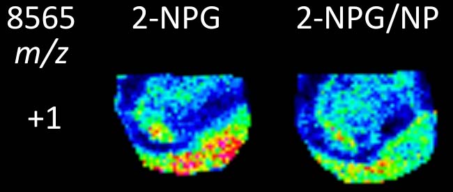
Results: The combined 2‐NPG and nanoparticle matrix produced highly charged ions from tissue with high‐vacuum MALDI. Nanoparticles of 20, 70, 400, and 1000 nm in diameter were tested, the 20 nm particles producing the highest charge states. Images of mouse brain tissue obtained from highly charged ions show similar spatial localization.
Conclusions: The combined 2‐NPG and nanoparticle matrix produces highly charged ions from tissue through a mechanism that may rely on the high surface area of the particles which can dry the tissue, and their ability to bind analyte molecules thereby assisting in crystal formation and production of multiply charged ions on laser irradiation.
